Abstract
Background:
Benign fibro-osseous lesions are characterized by the replacement of normal bone with cellular fibrous connective tissue with new bone formation. The published cytogenetic information on these tumors is limited to only few cases. Here, we report the cytogenetic and molecular genetic findings of a fibro-osseous tumor.
Methods:
A fibro-osseous lesion was investigated for genetic abnormalities using banding cytogenetics, fluorescence in situ hybridization (FISH), RNA sequencing, and direct cycle Sanger sequencing.
Results:
The karyotype was 46,XX,t(4;11;14;12)(q35;p15;q22;q13)[7]/46,XX [3], with no rearrangement of HMGA2. RNA sequencing revealed two FRMD6::PTH chimeric transcripts, originating from the fusion point 14q22;11p15 of the t(4;11;14;12). In these transcripts, exon 1 of FRMD6 fused to either exon 1 or exon 2 of PTH. Direct cycle sequencing confirmed the presence of these FRMD6::PTH chimeric transcripts.
Conclusion:
This study reports, for the first time, the presence of the FRMD6::PTH chimera in fibro-osseous tumor. In this chimera the expression of the entire coding region of PTH is regulated by the ubiquitously expressed FRMD6 gene promoter. Dysregulation of PTH expression may have significant implications for processes regulated by PTH protein.
Introduction
Benign fibro-osseous tumors of bone are neoplasms in which normal bone is replaced by cellular fibrous connective tissue with areas of new bone formation within the fibrous stroma [1, 2]. They are among the most common benign bone lesions in children and adolescents and represent a group of clinically distinct entities with significant histologic overlap [1, 2]. Although the exact cause of these tumors is unknown, they are believed to result from disruptions in the normal process of bone formation and remodeling [1, 2]. Fibro-osseous tumors occur in various body locations, including the skull, jawbones, and long bones, and typically exhibit slow growth over time. They encompass several types such as fibrous dysplasia, ossifying fibroma, osteo-fibrous dysplasia, and cemento-osseous dysplasia, which can lead to diagnostic challenges due to overlapping clinical, radiological, and pathological features [1, 2].
Published cytogenetic information on benign fibro-osseous tumors of bone is limited to few cases only [3, 4]. However, recent studies have suggested that diagnostic genetics may serve as an ancillary use in evaluating these tumors [1, 2]. Molecular genetic investigations have shown that fibro-osseous tumors can be associated with specific genetic alterations, which may aid in differential diagnosis. For example, in fibrous dysplasia somatic variants at codon 201 or codon 227 of the GNAS complex locus (GNAS on chromosomal sub-band 20q13.32) have been reported with frequencies ranging from 45% to 100% [5, 6]. These variants substitute arginine at codon 201 (p.R201) or glutamine at position 227 (p.Q227) resulting in constitutive activation of GNAS protein [5, 6]. Similarly, psammomatoid ossifying fibroma has been associated with rearrangements of the SATB homeobox 2 gene (SATB2 on chromosomal sub-band 2q33.1), as identified through whole transcriptome sequencing [7]. Moreover, a combination of whole exome sequencing and RNA sequencing has revealed fusion transcripts and variants in many genes in cemento-ossifying fibroma [8].
In the present study we applied a sequential approach combining karyotyping and RNA-sequencing to investigate a fibro-osseous tumor. Our aim was to identify neoplasm-specific fusion genes that could contribute to its pathogenesis.
Case presentation
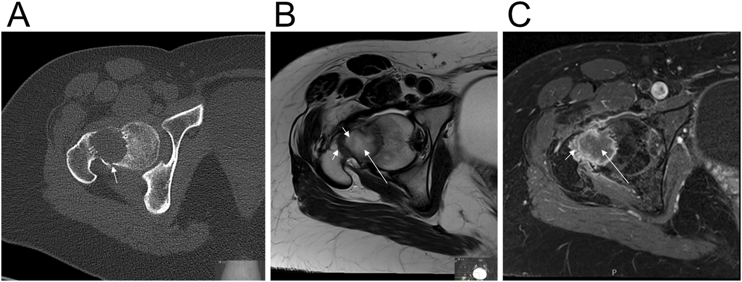
A 23-year-old woman presented with a 6-month history of pain in her right hip. The pain significantly worsened after minor trauma to the hip. She required the use of crutches and was admitted to a local hospital, where X-ray, computed tomography (CT), and magnetic resonance imaging (MRI) were performed. There were no previous imaging studies available for comparison. X-ray and CT revealed a well-defined osteolytic lesion in the femoral neck with a pathological fracture and no visible mineralization (Figure 1A). MRI showed that the lesion had a central area with high T2 signal intensity and high diffusion but no contrast enhancement, suggesting water content (Figures 1B,C). The lesion also had a peripheral border and intralesional stripes, both showing low T2 signal, which may represent fibrous content (Figure 1B). Contrast enhancement was seen along the peripheral border, indicating vascularized tissue (Figure 1C). Radiologically, the lesion had a benign appearance. Based on its location and signal pattern, cystic fibrous dysplasia or a simple bone cyst was suspected. The patient was treated with curettage of the lesion, packing with allogenic bone graft, and plate osteosynthesis.
FIGURE 1

Radiological features of the fibro-osseous tumor. (A) Axial CT image shows a well-defined osteolytic lesion in the femoral neck with a pathological fracture (arrow) and no visible mineralization. (B) Axial T2-weighted MRI of the lesion shows a central part with high signal intensity which may represent water content (long arrow), and a peripheral border as well as intralesional stripes, both with low signal intensity, which may correspond to fibrous content (short arrows). (C) Axial Fat-suppressed contrast-enhanced T1-weighted MRI of the lesion reveals that the central part doesn’t have contrast enhancement which representing water content (long arrow). The peripheral border has contrast enhancement indicating vascularized tissue (short arrow).
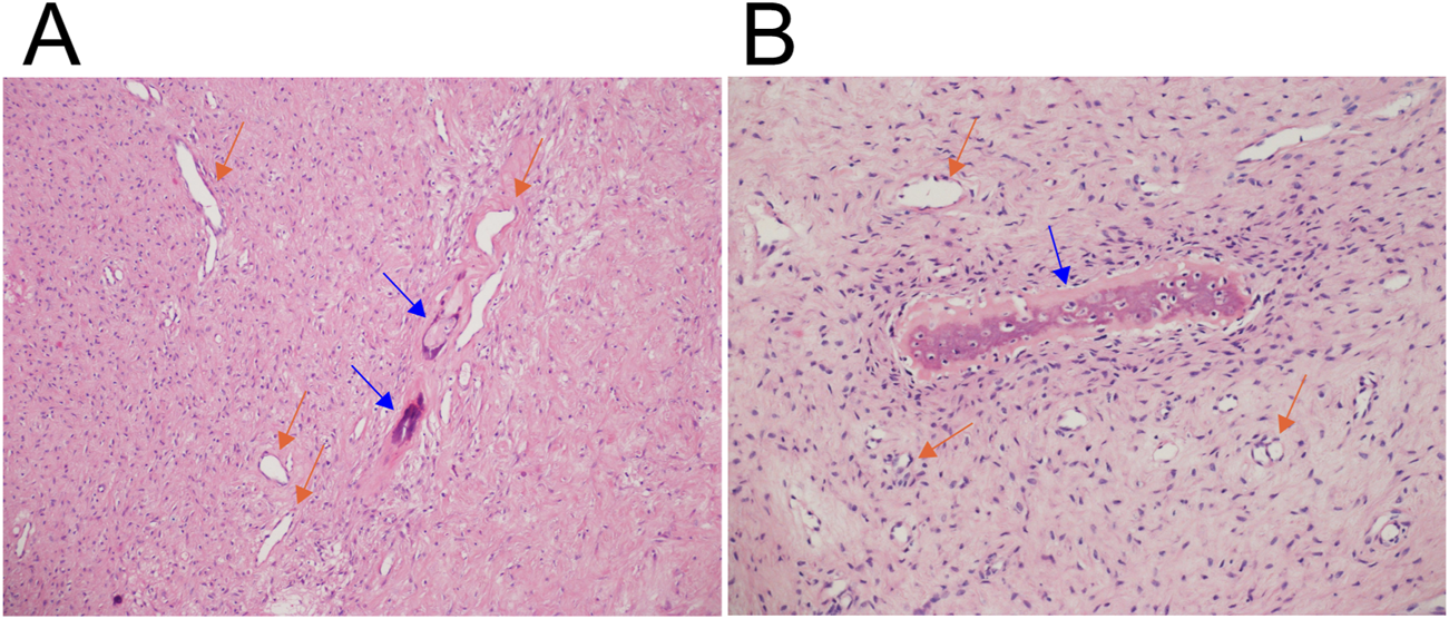
Microscopic examination revealed a lesion composed of both fibrous and osseous components (Figures 2A,B). The fibrous tissue lacked cytological atypia and was partially loosely organized. The osseous component consisted of scattered foci of new bone formation, lacking osteoblastic rimming. Immunohistochemical analysis demonstrated negative staining for S100 protein, desmin, ACTA2 (also known as alpha-smooth muscle actin, α-SMA), and CD34 (data not shown). The findings were consistent with a benign fibro-osseous lesion, although a more specific classification was not established.
FIGURE 2

Microscopic examination of the fibro-osseous tumor. Hematoxylin and eosin-stained sections show fibrous tissue lacking cytological atypia, with rich vascularization (orange arrows), and areas of loosely organized stroma. Scattered foci of new bone formation (blue arrows), lacking osteoblastic rimming, are also observed. (A) Magnification ×100. (B) Magnification ×200.
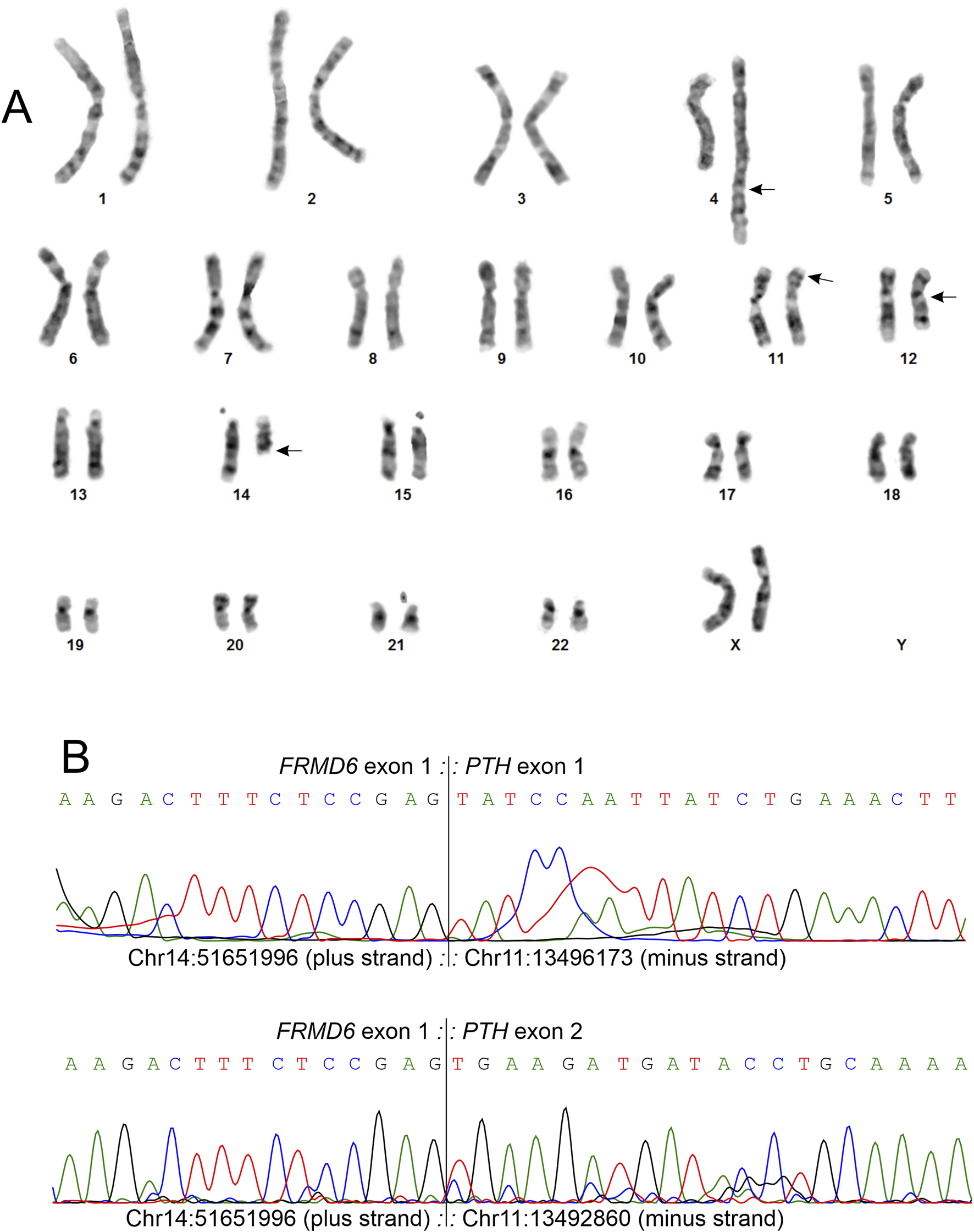
G-banding analysis of tumor cells [9] detected a four-way translocation involving chromosomal bands 4q35, 11p15, 14q22, and 12q13 as the sole cytogenetic aberration in 7 out of 10 examined metaphases (Figure 3A). The resulting abnormal karyotype was 46,XX,t(4;11;14;12)(q35;p15;q22;q13)[7]/46,XX[3]. FISH analysis on 96 interphase nuclei, using an in-house prepared HMGA2 break-apart probe as previously described [10], did not detected rearrangement of the HMGA2 gene (data not shown).
FIGURE 3

Genetic examination of the fibro-osseous tumor. (A) Karyogram showing the aberrant chromosomes 4, 11, 12, and 14 involved in a complex four-way translocation t(4;11;14;12)(q35;p15;q22;q13), along with their corresponding normal homologs. Breakpoint positions are indicated by arrows. (B) Partial Sanger sequencing chromatograms demonstrating the FRMD6::PTH fusion point, where exon 1 of the FRMD6 gene (transcript variant 2, NM_152330.4) is fused to exons 1 and 2 of the PTH gene (transcript variant 2, NM_001316352.2). The exact fusion point coordinates, based on the GRCh38/hg38 assembly, are chr14: 51651996 (plus strand) for FRMD6 exon 1 and chr11:13496173 and for chr11:13492860 (both minus strand) for PTH exons 1 and 2, respectively. The two fusion transcript sequences have been submitted to the GenBank database and assigned the accession numbers PV085792 and PV085793.
Total RNA extracted from frozen (−80°C) tumor tissue adjacent to that used for cytogenetic and histological analyses underwent high-throughput paired-end sequencing at the Genomics Core Facility, Norwegian Radium Hospital, Oslo University Hospital. Using FusionCatcher software [11] with fastq files from RNA sequencing, two FRMD6::PTH chimeric transcript sequences were detected, corresponding to the fusion points 14q22; 11p15 of the aforementioned four-way translocation. In the first transcript, exon 1 of FRMD6 fused to exon 1 of PTH: 5′-AGA GGG GTG ACC AGA GAG CCC AAC GCC TGG TGC TCA AGA CTT TCT CCG AG::TA TCC AAT TAT CTG AAA CTT AAG AAG AGT GTG CAC CGC CCA ATG GGT GTG-3´. In the second FRMD6::PTH chimeric transcript, exon 1 of FRMD6 fused to exon 2 of PTH: 5′-GTG ACC AGA GAG CCC AAC GCC TGG TGC TCA AGA CTT TCT CCG AG::TG AAG ATG ATA CCT GCA AAA GAC ATG GCT AAA GTT ATG ATT GTC-3′.
To confirm the presence of the fusion transcripts, complementary DNA (cDNA) was synthesized from 400 ng of total RNA and cDNA corresponding to 20 ng of total RNA was used as the template in the subsequent PCR/cycle (Sanger) assays using the BigDye Direct Cycle Sequencing Kit, following the manufacturer’s recommendations (ThermoFisher Scientific, Waltham, MA, United States). The primer combinations were FRMD6-13F1 (5′-AGG CTC GGC GCC GGT AGG AA-3′)/PTH-40R1 (5′-CAC ACA CCC ATT GGG CGG TGC-3′) and FRMD6-26F1 (5′- GTA GGA AGA GTC AGA GGG GTG ACC A-3′)/PTH-204R1 (5′-AAC AGA TTT CCC ATC CGA TTT TGT AA-3′). The FRMD6 forward primers had the M13 forward primer sequence (TGTAAAACGACGGCCAGT) at their 5′-end, and the PTH reverse primers had the M13 reverse primer sequence (CAGGAAACAGCTATGACC) at their 5′-end. Sequence analysis was performed using the Applied Biosystems SeqStudio Genetic Analyzer (ThermoFisher Scientific), and the obtained sequencing data were aligned with the reference sequences NM_152330.4 for FRMD6 and NM_001316352.2 for PTH using the Basic Local Alignment Search Tool (BLAST). Direct cycle sequencing confirmed the presence of both FRMD6::PTH transcripts, as shown in Figure 3B. The two fusion transcript sequences have been submitted to the GenBank database and have been assigned the accession numbers PV085792 and PV085793. The Supplementary Data Sheet provides detailed descriptions of the methodologies used for G-banding and karyotyping, fluorescence in situ hybridization (FISH), RNA sequencing, and molecular genetic analyses.
Discussion
The FRMD6 gene (located at sub-band 14q22.1) encodes the 4.1-ezrin-radixin-moesin (FERM) domain-containing protein 6 (FRMD6, also known as Willin) [12, 13]. This protein has diverse cellular functions, including the regulation of the Hippo and mTOR signaling pathways [13]. FRMD6 has four transcript variants (accession numbers NM_001042481.3, NM_001267046.2, NM_001267047.1, and NM_152330.4) and is broadly expressed across multiple tissues [12, 13]. In these transcripts, either exon 1 (NM_001267046.2, NM_001267047.1, and NM_152330.4) or exons 1 and 2 (NM_001042481.3) are untranslated. Chimeras involving FRMD6 as the 5′-end partner gene have been reported in less than ten tumors [14, 15] (Table 1). In all these cases, the untranslated exons of FRMD6 fused to the 3′-end partner gene, with the promoter and other 5′-end regulatory elements of FRMD6 driving the expression of the 3′-end partner gene [16, 17].
TABLE 1
| Tumor type | Chimera | Reference |
|---|---|---|
| head and neck squamous cell carcinoma | FRMD6 [NM_001042481.3] exon 2::BPIFB1 [NM_033197.3] exon 2 | [14] |
| mesothelioma | FRMD6 [NM_152330.4] exon 1::GNG2 [NM_053064.5] exon 3 | [14] |
| bladder urothelial carcinoma | FRMD6 [NM_001042481.3] exon 2::RTN4 [NM_007008.3] exon 2 | [14] |
| lung adenocarcinoma | FRMD6 [NM_152330.4] exon 1::SCFD1 [NM_016106.4] exon 15 | [14] |
| salivary duct carcinoma | FRMD6 [NM_152330.4] exon 1::PLAG1 [NM_002655.2] exon 3 | [15] |
| lung squamous cell carcinoma | BTBD10 [NM_032320.7] exon 1::PTH [NM_001316352.1] exon 1 | [14] |
| breast invasive carcinoma | NUMA1 [NM_006185.4] exon 1::PTH [NM_001316352.1] exon 1 | [14] |
| fibro-osseous tumor |
FRMD6 [NM_152330.4] exon 1::PTH [NM_001316352.2] exon 1 FRMD6 [NM_152330.4] exon 1::PTH [NM_001316352.2] exon 2 |
Present study |
Published cases in which chimeric genes have FRMD6 as the 5′-end partner and cases in which chimeric genes have PTH as 3′-end partner.
The PTH gene (located at sub-band 11p15.2) is exclusively expressed in the parathyroid glands, where it encodes parathyroid hormone (PTH) [18, 19]. This hormone plays a crucial role in regulating blood calcium levels, as well as in phosphorus metabolism, bone formation, and resorption [18, 19]. The expression of the PTH gene is regulated by numerous transcription factors, including both activators and repressors [20, 21]. A 5.2-kbp DNA region immediately upstream of the human PTH gene was found to be sufficient in order to drive parathyroid-specific gene expression [21]. Ectopic expression of PTH by tumor cells has been reported in fewer than 30 cases, often contributing to hypercalcemia associated with malignancy [22]. In an ovarian carcinoma the expression was linked to rearrangement of the PTH gene [23], while in a case of high-grade neuroendocrine carcinoma of the pancreas, ectopic PTH expression was associated with hypomethylation of the PTH locus [24]. In other non-parathyroid tumors, however, ectopic PTH expression was not associated with rearrangement, amplification or hypomethylation of the PTH gene [22]. Chimeric transcripts with PTH as the 3′-end partner gene have been described in two tumors: in a lung squamous cell carcinoma, where exon 1 of the BTB domain containing 10 (BTBD10) gene fused to exon 1 of PTH (BTBD10::PTH) and in a breast invasive carcinoma, where exon 1 of the nuclear mitotic apparatus protein 1 (NUMA1) fused to exon 1 of PTH (NUMA1::PTH) [14]. These chimeric transcripts indicate genomic rearrangements involving both PTH and the respective 5′-end partner genes, suggesting that the promoters and other 5′-end regulatory elements of the ubiquitously expressed BTBD10 or NUMA1 control PTH expression.
To the best of our knowledge, the FRMD6::PTH chimera, identified in the examined fibro-osseous tumor, is reported for the first time in this study. The FRMD6::PTH chimera is predicted to be located on the der (14) chromosome resulting from the four-way translocation, as both the PTH gene (located on 11p15.2) and the FRMD6 gene (located on 14q22.1) are transcribed from centromere to telomere. The FRMD6::PTH chimeric transcript follows a pattern observed in previous reported chimeras involving FRMD6 as the 5′-end partner gene, where the untranslated exons of FRMD6 fused to the 3′-end partner gene. It also exhibits the pattern found in the two previously described chimeric transcripts with PTH as the 3′-end partner gene, involving the fusion of the entire translated part of PTH, with its expression controlled by the promoter and other 5′-end regulatory elements of ubiquitously expressed genes. Dysregulation of PTH expression may have implications for calcium homeostasis and other processes regulated by PTH protein.
Although the presence of the chimeric transcript strongly suggests transcriptional activation of PTH in the tumor, the actual expression of functional PTH protein and activation of downstream signaling pathways remain speculative. Potential regulatory mechanisms may include promoter swapping, whereby the promoter of FRMD6 -a gene expressed in a wide range of tissues-drives ectopic expression of PTH, as well as changes in chromatin structure or local epigenetic alterations induced by the translocation event [25, 26]. Moreover, the preservation of the full PTH coding sequence in the chimera raises the possibility that biologically active hormone could be produced, although this has not yet been demonstrated.
Importantly, PTH is not only a regulator of calcium metabolism but also plays a role in bone remodeling by stimulating both osteoclastic bone resorption and osteoblastic bone formation [27, 28]. During these remodeling processes, fibrous tissue may form as part of the bone repair response, especially in areas undergoing resorption. PTH has also been shown to enhance vascularization within bone by promoting microvessel formation [29], which may support the development of a fibrous stroma. These biological effects suggest that aberrant PTH expression in tumor cells could contribute to the fibro-osseous characteristics of the tumor, potentially by promoting fibrous matrix production, increased vascularity, and altered bone metabolism.
Future functional studies are needed to explore the biological significance of this fusion. These should include analyses at the transcript and protein levels to confirm PTH expression, for example using qRT-PCR, western blotting, or immunohistochemistry. Assays to evaluate PTH1R-mediated signaling, such as measurement of cAMP accumulation or phosphorylation of downstream effectors, could help determine whether the PTH pathway is functionally active [30, 31]. In vitro expression of the FRMD6::PTH fusion construct in model cell lines would provide further insight into its biological role and could clarify whether the fusion promotes tumor cell proliferation, survival, osteogenic differentiation, or fibrous tissue formation, as has been shown for other oncogenic fusion genes [32]. Such investigations would provide valuable insights into whether this fusion plays a direct oncogenic role or represents a passenger alteration.
Conclusion
This study reports the first fibro-osseous tumor carrying the FRMD6::PTH chimeric gene, resulting from a chromosomal aberration. This fusion potentially disrupts PTH expression, warranting further investigation into its role in tumorigenesis.
Statements
Data availability statement
The original contributions presented in the study are included in the article/Supplementary Material, further inquiries can be directed to the corresponding author.
Ethics statement
The studies involving humans were approved by Regional Committee for Medical and Health Research Ethics, South-East Norway (Regional komité for medisinsk forskningsetikk Sør-Øst, Norge; http://helseforskning.etikkom.no). The studies were conducted in accordance with the local legislation and institutional requirements. The participants provided their written informed consent to participate in this study. Written informed consent was obtained from the individual(s) for the publication of any potentially identifiable images or data included in this article.
Author contributions
IP conceived the molecular experiments and analyzed data. KA performed molecular and FISH investigations. IsL performed the radiological examination. LG perform the cytogenetic investigation. InL performed histopathological and immunohistochemical examinations. IP takes full responsibility for the work as a whole, including the study design, access to data, and the decision to submit and publish the manuscript. All authors contributed to the article and approved the submitted version.
Funding
The author(s) declare that no financial support was received for the research and/or publication of this article.
Conflict of interest
The authors declare that the research was conducted in the absence of any commercial or financial relationships that could be construed as a potential conflict of interest.
Generative AI statement
The author(s) declare that no Generative AI was used in the creation of this manuscript.
Supplementary material
The Supplementary Material for this article can be found online at: https://www.por-journal.com/articles/10.3389/pore.2025.1612096/full#supplementary-material
References
1.
Velez Torres JM Rosenberg AE . Benign fibro-osseous tumors of bone: clinicopathological findings and differential diagnosis. Diagn Histopathology (2022) 28(12):510–21. 10.1016/j.mpdhp.2022.09.002
2.
Chebib I Chang CY Lozano-Calderon S . Fibrous and fibro-osseous lesions of bone. Surg Pathol Clin (2021) 14(4):707–21. 10.1016/j.path.2021.06.011
3.
Dal Cin P Sciot R Brys P De Wever I Dorfman H Fletcher CD et al Recurrent chromosome aberrations in fibrous dysplasia of the bone: a report of the CHAMP study group. CHromosomes and MorPhology. Cancer Genet Cytogenet (2000) 122(1):30–2. 10.1016/s0165-4608(00)00270-3
4.
Parham DM Bridge JA Lukacs JL Ding Y Tryka AF Sawyer JR . Cytogenetic distinction among benign fibro-osseous lesions of bone in children and adolescents: value of karyotypic findings in differential diagnosis. Pediatr Dev Pathol (2004) 7(2):148–58. 10.1007/s10024-003-6065-z
5.
Lee SE Lee EH Park H Sung JY Lee HW Kang SY et al The diagnostic utility of the GNAS mutation in patients with fibrous dysplasia: meta-analysis of 168 sporadic cases. Hum Pathol (2012) 43(8):1234–42. 10.1016/j.humpath.2011.09.012
6.
Tabareau-Delalande F Collin C Gomez-Brouchet A Decouvelaere AV Bouvier C Larousserie F et al Diagnostic value of investigating GNAS mutations in fibro-osseous lesions: a retrospective study of 91 cases of fibrous dysplasia and 40 other fibro-osseous lesions. Mod Pathol (2013) 26(7):911–21. 10.1038/modpathol.2012.223
7.
Cleven AHG Szuhai K van IDGP Groen E Baelde H Schreuder WH et al Psammomatoid ossifying fibroma is defined by SATB2 rearrangement. Mod Pathol (2023) 36(1):100013. 10.1016/j.modpat.2022.100013
8.
Gomez RS El Mouatani A Duarte-Andrade FF Pereira T Guimaraes LM Gayden T et al Comprehensive genomic analysis of cemento-ossifying fibroma. Mod Pathol (2023) 37(2):100388. 10.1016/j.modpat.2023.100388
9.
Lukeis R Suter M . Cytogenetics of solid tumours. Methods Mol Biol (2011) 730:173–87. 10.1007/978-1-61779-074-4_13
10.
Panagopoulos I Gorunova L Andersen K Lund-Iversen M Hognestad HR Lobmaier I et al Chromosomal translocation t(5;12)(p13;q14) leading to fusion of high-mobility group at-hook 2 gene with intergenic sequences from chromosome sub-band 5p13.2 in benign myoid neoplasms of the breast: a second case. Cancer Genomics Proteomics (2022) 19(4):445–55. 10.21873/cgp.20331
11.
Kangaspeska S Hultsch S Edgren H Nicorici D Murumagi A Kallioniemi O . Reanalysis of RNA-sequencing data reveals several additional fusion genes with multiple isoforms. PLoS One (2012) 7(10):e48745. 10.1371/journal.pone.0048745
12.
Gunn-Moore FJ Welsh GI Herron LR Brannigan F Venkateswarlu K Gillespie S et al A novel 4.1 ezrin radixin moesin (FERM)-containing protein, 'Willin. FEBS Lett (2005) 579(22):5089–94. 10.1016/j.febslet.2005.07.097
13.
Chen D Yu W Aitken L Gunn-Moore F . Willin/FRMD6: a Multi-functional neuronal protein associated with Alzheimer's disease. Cells (2021) 10(11):3024. 10.3390/cells10113024
14.
Gao Q Liang WW Foltz SM Mutharasu G Jayasinghe RG Cao S et al Driver fusions and their implications in the development and treatment of human cancers. Cell Rep (2018) 23(1):227–38.e3. 10.1016/j.celrep.2018.03.050
15.
Lassche G van Helvert S Eijkelenboom A Tjan MJH Jansen EAM van Cleef PHJ et al Identification of fusion genes and targets for genetically matched therapies in a large cohort of salivary gland cancer patients. Cancers (Basel) (2022) 14(17):4156. 10.3390/cancers14174156
16.
Renz PF Valdivia-Francia F Sendoel A . Some like it translated: small ORFs in the 5'UTR. Exp Cell Res (2020) 396(1):112229. 10.1016/j.yexcr.2020.112229
17.
Ryczek N Lys A Makalowska I . The Functional meaning of 5'UTR in protein-coding genes. Int J Mol Sci (2023) 24(3):2976. 10.3390/ijms24032976
18.
Silva BC Bilezikian JP . Parathyroid hormone: anabolic and catabolic actions on the skeleton. Curr Opin Pharmacol (2015) 22:41–50. 10.1016/j.coph.2015.03.005
19.
Liu H Liu L Rosen CJ . PTH and the regulation of mesenchymal cells within the bone marrow niche. Cells (2024) 13(5):406. 10.3390/cells13050406
20.
Koszewski NJ Alimov AP Langub MC Park-Sarge OK Malluche HH . Contrasting mammalian PTH promoters: identification of transcription factors controlling PTH gene expression. Clin Nephrol (2005) 63(2):158–62. 10.5414/cnp63158
21.
Mallya SM Wu HI Saria EA Corrado KR Arnold A . Tissue-specific regulatory regions of the PTH gene localized by novel chromosome 11 rearrangement breakpoints in a parathyroid adenoma. J Bone Miner Res (2010) 25(12):2606–12. 10.1002/jbmr.187
22.
Uchida K Tanaka Y Ichikawa H Watanabe M Mitani S Morita K et al An excess of CYP24A1, lack of CaSR, and a novel lncRNA near the PTH gene characterize an ectopic PTH-producing tumor. J Endocr Soc (2017) 1(6):691–711. 10.1210/js.2017-00063
23.
Nussbaum SR Gaz RD Arnold A . Hypercalcemia and ectopic secretion of parathyroid hormone by an ovarian carcinoma with rearrangement of the gene for parathyroid hormone. N Engl J Med (1990) 323(19):1324–8. 10.1056/NEJM199011083231907
24.
VanHouten JN Yu N Rimm D Dotto J Arnold A Wysolmerski JJ et al Hypercalcemia of malignancy due to ectopic transactivation of the parathyroid hormone gene. J Clin Endocrinol Metab (2006) 91(2):580–3. 10.1210/jc.2005-2095
25.
Esteller M Dawson MA Kadoch C Rassool FV Jones PA Baylin SB . The Epigenetic hallmarks of cancer. Cancer Discov (2024) 14(10):1783–809. 10.1158/2159-8290.CD-24-0296
26.
Lavaud M Tesfaye R Lassous L Brounais B Baud'huin M Verrecchia F et al Super-enhancers: drivers of cells' identities and cells' debacles. Epigenomics (2024) 16(9):681–700. 10.2217/epi-2023-0409
27.
Wojda SJ Donahue SW . Parathyroid hormone for bone regeneration. J Orthop Res (2018) 36(10):2586–94. 10.1002/jor.24075
28.
Mehreen A Faisal M Zulfiqar B Hays D Dhananjaya K Yaseen F et al Connecting bone remodeling and regeneration: unraveling hormones and signaling pathways. Biology (Basel) (2025) 14(3):274. 10.3390/biology14030274
29.
Menger MM Tobias AL Bauer D Bleimehl M Scheuer C Menger MD et al Parathyroid hormone stimulates bone regeneration in an atrophic non-union model in aged mice. J Transl Med (2023) 21(1):844. 10.1186/s12967-023-04661-y
30.
Goltzman D . Physiology of parathyroid hormone. Endocrinol Metab Clin North Am (2018) 47(4):743–58. 10.1016/j.ecl.2018.07.003
31.
Martin TJ . PTH1R actions on bone using the cAMP/protein kinase A pathway. Front Endocrinol (Lausanne) (2021) 12:833221. 10.3389/fendo.2021.833221
32.
Chang Y Zhao Z Song Y . Research progress on fusion genes in tumours. Clin Translational Discov (2024) 4(4):e352. 10.1002/ctd2.352
Summary
Keywords
fibro-osseous tumor, chromosomal translocation, FERM domain containing 6 (FRMD6), parathyroid hormone (PTH), FRMD6::PTH chimera
Citation
Panagopoulos I, Andersen K, Lloret I, Gorunova L and Lobmaier I (2025) Novel FRMD6::PTH chimera in tumorous bone lesion carrying a t(4;11;14;12)(q35;p15;q22;q13). Pathol. Oncol. Res. 31:1612096. doi: 10.3389/pore.2025.1612096
Received
31 January 2025
Accepted
16 June 2025
Published
26 June 2025
Volume
31 - 2025
Edited by
Anna Sebestyén, Semmelweis University, Hungary
Updates
Copyright
© 2025 Panagopoulos, Andersen, Lloret, Gorunova and Lobmaier.
This is an open-access article distributed under the terms of the Creative Commons Attribution License (CC BY). The use, distribution or reproduction in other forums is permitted, provided the original author(s) and the copyright owner(s) are credited and that the original publication in this journal is cited, in accordance with accepted academic practice. No use, distribution or reproduction is permitted which does not comply with these terms.
*Correspondence: Ioannis Panagopoulos, ioapan@ous-hf.no
Disclaimer
All claims expressed in this article are solely those of the authors and do not necessarily represent those of their affiliated organizations, or those of the publisher, the editors and the reviewers. Any product that may be evaluated in this article or claim that may be made by its manufacturer is not guaranteed or endorsed by the publisher.